|
Important Landmarks
|
Sonographic Images
|
|
olecranon
|
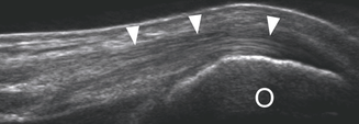
Figure 42. This is a sonographic image of the inertion of the triceps brachii on the olecranon of the ulna. O-olecranon, white arrows-pointing to the triceps brachii tendon. Adapted from "Lecture 6 Ultrasound of the Elbow" by Laura Thomas, 2014.
|
|
cubital tunnel
|
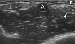
Figure 43. This is a sonographic image of the cubital tunnel (anterior compartment of the elbow). T-triceps brachii tendon, A-brachial artery, white arrows- pointing to the median nerve. Adapted from "Lecture 6 Ultrasound of the Elbow" by Laura Thomas, 2014.
|
|
ulnar nerve
|
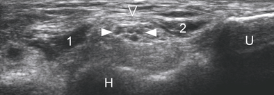
Figure 44. This is a sonographic image of the ulnar nerve. U-ulna, H-humerus, 1/2-both heads of the flexor carpi ulnaris muscle, white arrows-pointing to the ulnar nerve. Adapted from "Lecture 6 Ultrasound of the Elbow" by Laura Thomas, 2014.
|
|
biceps brachii
(biceps tendon) |
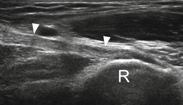
Figure 45. This is a sonographic image of the biceps brachii tendon insertion onto the radial tuberosity. R-radius, white arrows-pointing to the biceps brachii tendon. Adapted from "Lecture 6 Ultrasound of the Elbow" by Laura Thomas, 2014.
|
|
annular ligament
|
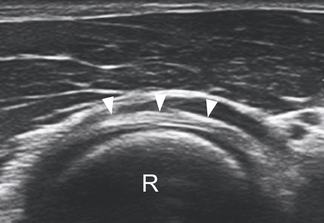
Figure 46. This is a sonographic image of the annular ligament surrounding the head of the radius. R-radius, white arrows-annular ligament. Adapted from "Lecture 6 Ultrasound of the Elbow" by Laura Thomas, 2014.
|
