|
Pathology
|
Description
|
Sonographic Findings
|
Sonographic Images
|
|
DeQuervain (sclerosing) tenosynovitis
|
-differs from the more common variants of tenosynovitis in that there is often less fluid and more tenosynovial thickening
-affects the first extensor compartment |
-distention of the tendon sheath with fluid, hypoechoic synovial proliferation, hyperaemic changes within the tendon sheath may be evident, but the tendon itself stays relatively the same in its echotexture
|
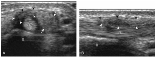
Figure 59. This is a sonographic image demonstrating DeQuervians disease. Image A is short axis of the first extensor wrist tendons and image B is the long axis of the first extensor wrist tendons. Arrows-pointing to hypoechoic swelling of the abductor pollicis longus, arrowheads-pointing to the hypoechoic thickening of the tendon sheath, E-extensor pollicis brevis tendon, R-radius. Adapted from "Lecture 10 Wrist Pathology (2)" by Laura Thomas.
|
|
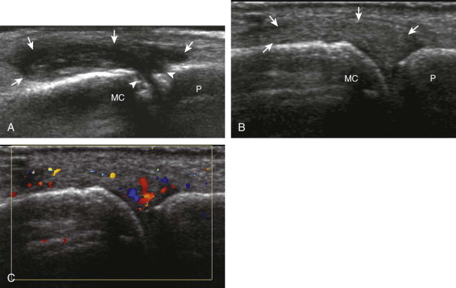
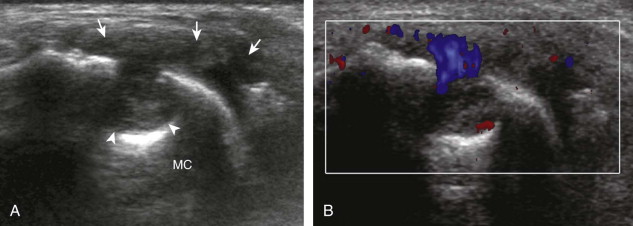
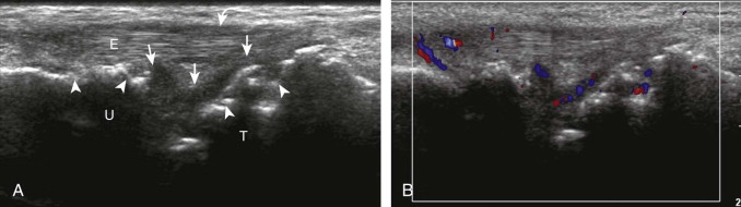
rheumatiod arthritis
|
-a chronic, inflammatory, destructive an sometimes deforming collagen disease that has an autoimmune component
-it is characterized by symmetric inflammation of synovial membranes and increased synovial exudate, leading to thickening of the membranes and swelling of the joints |
-synovial hypertrophy appearing as hypoechoic (most commonly), isoechoic or hyperechoic relative to the subdermal fat, poorly compressible tissue within the joint space
-flow may be demonstrated with colour or power doppler, depending on the inflammatory activity -joint synovial hypertrophy may be seen in the dorsal recesses of the wrist, the volar and dorsal recesses of the metacarpophalangeal and interphalangeal joints of the hand -erosions appear as discontinuity of the bone cortex seen in two orthogonal planes and these erosions begin in the marginal regions of a joint, where the bone cortex is not covered with hyaline cartilage and is directly exposed to joint inflammation (the finding of synovial hypertrophy directly over a cortical irregularity also increases the likelihood that an erosion is present) |
|
(Jacobson, 2007)
(McNally, 2005, pg. 95, 98-99)
(McNally, 2005, pg. 95, 98-99)
