|
Important Landmarks
|
Images
|
|
wrist compartment 1
|
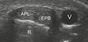
Figure 55. This is a sonographic image of wrist compartment 1. APL-abductor pollicis longus, EPB-extensor pollicis brevis, R-radius. Adapted from "Lecture 8 Forearm and Wrist Pres" by Laura Thomas, 2014.
|
|
wrist compartment 3
|
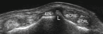
Figure 56. This is a sonographic image of compartment 3 of the wrist. EPL- extensor pollicis longus, ECRB-extensor carpi radialis brevis, ECRL-extensor carpi radialis longus. Adapted from "Lecture 8 Forearm and Wrist Pres" by Laura Thomas, 2014.
|
|
median nerve
|
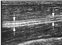
Figure 57. This is a sonographic image demonstrating the median nerve in long axis. Adapted from "Lecture 8 Forearm and Wrist Pres" by Laura Thomas, 2014.
|
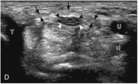
Figure 58. This is a sonographic image demonstrating the median nerve in short axis within the carpal tunnel.Black arrows-pointing to the retinaculum, white arrows-pointing to the median nerve. Adapted from "Fundamentals of Musculoskeletal Ultrasound" by Jacobson, A, Jon, 2007, Wrist and Hand Ultrasound, 5. Copyright by Elsevier, Inc.
|
